
Figure 8.
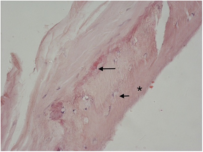
Area of talonavicular ligament insertion into bone (*) in which there are osteocyte nuclei (short arrow). There is a line of calcification (long arrow) at the point of insertion into bone above which there is loss of the parallel arrangement of collagen fibres in the ligament. Haematoxylin–eosin: original magnification ×400.