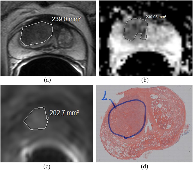
Figure 1.
Comparison of intersequence volumes with histology: transverse T2 weighted (T2W) (a), diffusion-weighted (DW-) (b) and dynamic contrast enhanced (DCE-) images at 30 s post-injection of gadoterate meglumine (c). Tumour outlines drawn on three separate occasions by Observer 1 are overlaid. The volume in (a) was largest, and the volume in (c) was smallest, although overlap between the outlines is noted in all cases. Whole-mount histological specimen at prostatectomy (d) confirms the presence of tumour at that location.

