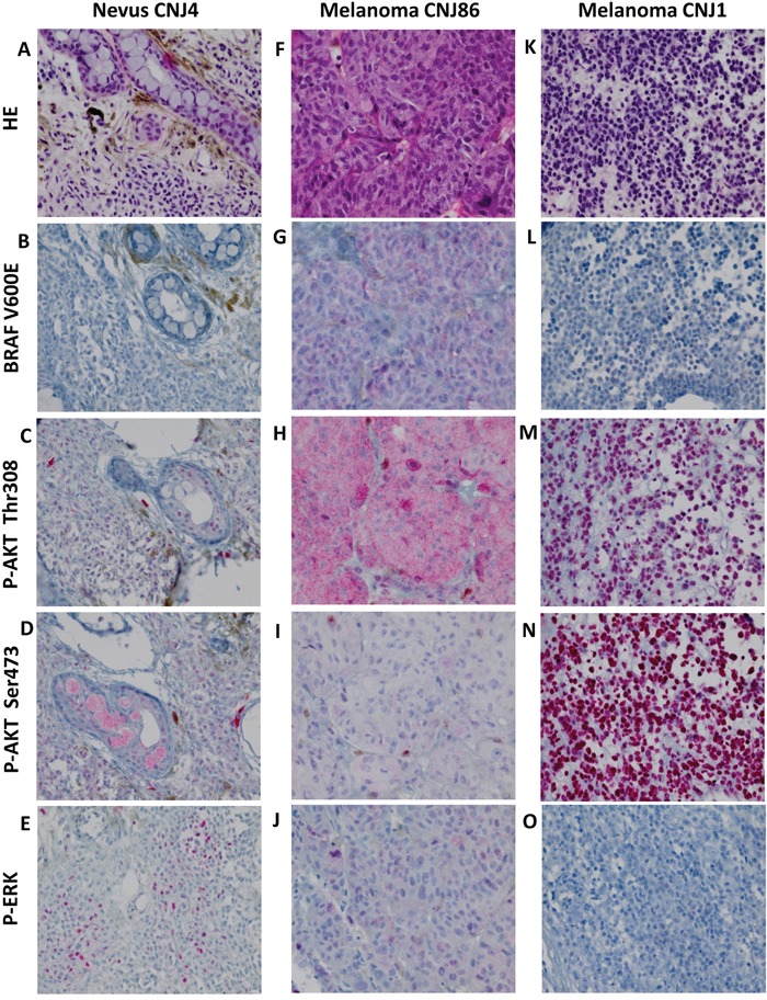
Figure 1. HE staining, BRAF V600E expression and phosphorylation of ERK and AKT in CM.

A-E. nevus, F-J. BRAF V600E mutated melanoma, K-O. BRAF V600E negative melanoma. HE staining (A, F, K). Positive staining for p-AKT Thr308, p-AKT Ser473 and p-ERK occurred in a cytoplasmic (H, I) and nuclear (C, M, N) fashion and in combination (D, E, J). Negative staining (B, L, O). Staining patterns for these proteins did not correlate with malignant progression. All images were taken at the magnification of ×400.
