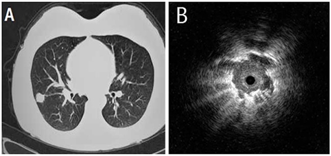
Figure 1. A 52-year-old male who underwent right middle lung lobectomy for pulmonary adenocarcinoma.

(A) Chest computed tomography showed a pulmonary nodule of 26 mm in diameter. (B) Endobronchial ultrasonography showed a low echoic nodule surrounded by a strong reflected interface produced between the aerated lung and the lesion.
