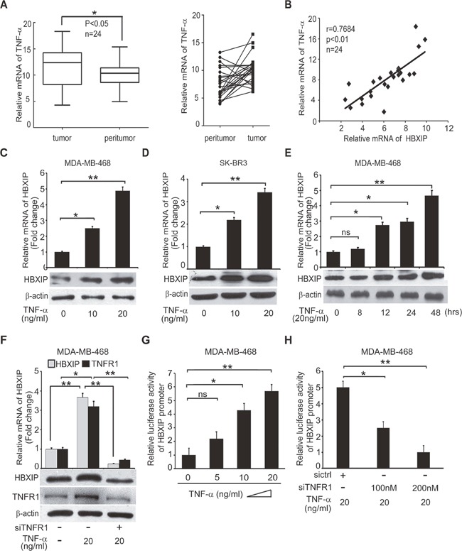
Figure 2. TNF-α is positively correlated with HBXIP in clinical breast cancer tissues and up-regulates HBXIP in breast cancer cells.

(A) The relative mRNA levels of TNF-α were detected by qRT-PCR in clinical breast cancer tissues (n=24) (*p<0.05, Wilcoxon's signed-rank test). (B) The relative mRNA levels of HBXIP and TNF-α were detected by qRT-PCR and normalized against GAPDH in above samples (Spearman's correlation = 0.7684, **p < 0.01). (C, D) The mRNA and protein levels of HBXIP were measured by qRT-PCR and Western blot analysis in MDA-MB-468 and SK-BR3 cells treated with 10 ng/ml, 20 ng/ml TNF-α, respectively. (E) The expression of HBXIP in mRNA and protein levels were measured by qRT-PCR and Western blot analysis in MDA-MB-468 cells treated with 20 ng/ml TNF-α in a time course (0-48 hours). (F) The expression levels of HBXIP and TNFR1 were measured by qRT-PCR and Western blot assays in MDA-MB-468 cells treated with 20 ng/ml TNF-α and transfected with siTNFR1. (G, H) Dual luciferase reporter gene assays were performed to detect the relative activities of HBXIP promoter in MDA-MB-468 cells added into 5 ng/ml, 10 ng/ml and 20 ng/ml TNF-α and/or transiently transfected with 100 nM, 200 nM siTNFR1. Error bars represent ±s.d., *p<0.05, **p< 0.01, Student's t test. All experiments were performed at least 3 times.
