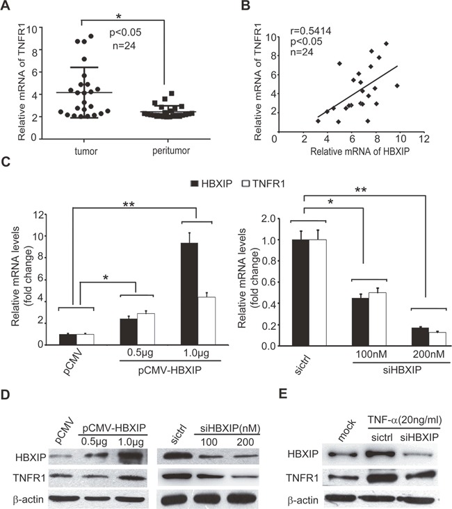
Figure 5. TNF-α-elevated HBXIP up-regulates the expression of TNFR1 in breast cancer cells.

(A) The expression levels of TNFR1 were detected by qRT-PCR analysis in clinical breast cancer tissues (n=24) (*p<0.05, Wilcoxon's signed-rank test). (B) The relative mRNA levels of HBXIP and TNFR1 were examined by qRT-PCR analysis in above samples (Spearman's correlation = 0.5414, *p < 0.05). (C) The relative fold changes of TNFR1 and HBXIP mRNA levels were detected by qRT-PCR analysis in MDA-MB-468 cells transiently transfected with pCMV-HBXIP or siHBXIP. (D, E) The expression levels of TNFR1 and HBXIP were examined by Western blot analysis in MDA-MB-468 cells transiently transfected with pCMV-HBXIP or siHBXIP and/or treated with 20 ng/ml TNF-α. Error bars represent ±s.d., *p < 0.05, **p < 0.01, Student's t test. All experiments were repeated at least 3 times.
