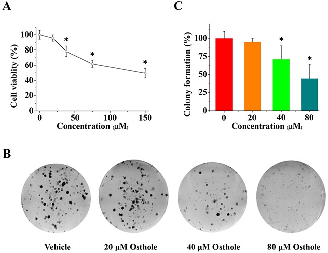
Figure 3. Osthole inhibited breast cancer cell viability and proliferation.

(A) Osthole treatment inhibited MDA-231BO cell viability. Cells were treated with vehicle or osthole (18.75, 37.5, 75 and 150 μM) for 24 h and then measured using MTT assays. Results are expressed as mean ± SD for three experiments (* p < 0.05 by ANOVA). (B) Representative images from colony formation assays using cells treated with either osthole or vehicle were taken at 24 h. (C) Quantitative results of colony formation (% of vehicle) expressed as the mean ± SD of three experiments. * p < 0.05 vs. vehicle. Colony formation of vehicle-treated cells was set at 100 %, and values for colony formation of osthole-treated cells were represented as a percentage of vehicle colony formation (* p < 0.05 by ANOVA).
