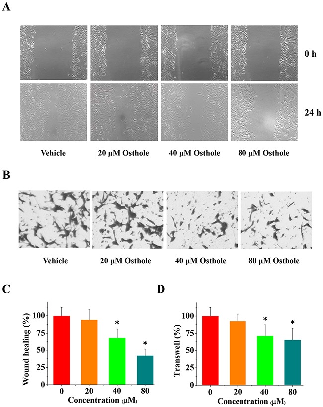
Figure 4. The anti-invasion and anti-migration effects of osthole on breast cancer cells.

(A) Representative images from wound healing assays using cells treated with either osthole or vehicle were taken at 0 h and 24 h. (B) Representative images of transwell analysis of cells treated with either osthole or vehicle were taken at 24 h. (C) Wound closure was quantified as the percentage of wound closure, and expressed as the mean ± SD of three experiments. Wound closure of vehicle-treated cells was set at 100 %, and wound closure of osthole-treated cells was represented as a percentage of the vehicle wound closure (* p < 0.05 by ANOVA). (D) Quantitative results of transwell migration assay (% of vehicle) expressed as the mean ± SD of three experiments. * p < 0.05 vs. vehicle. Transwell assay data, with vehicle-treated cells set at 100%, and osthole-treated cells represented as a percentage of the vehicle group (* p < 0.05 by ANOVA).
