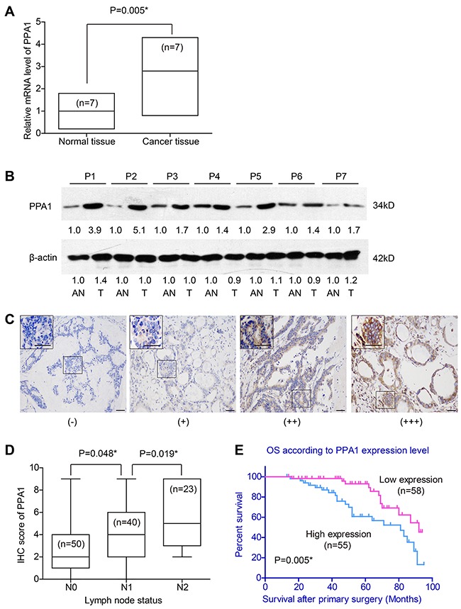
Figure 1. Expression of PPA1 in colon adenocarcinoma tissues.

(A) RT-qPCR results showed higher mRNA levels of PPA1 in cancer tissues than that in adjacent normal tissues (P=0.005). (B) Western Blot revealed the different expression levels of PPA1 protein in tumor tissues (T) and adjacent normal tissues (AN). The fold changes were labelled under the bands using AN as control. P1-P7 refer to the patient's number from whom we obtained the fresh-frozen tissues. (C) IHC of tumor tissues showed different immunoreactivities. Scale bar: 100μm. (D) Patients with advanced lymph node statues exhibited higher PPA1 levels as determined by IHC evaluation. (E) High expression of PPA1 indicated poorer overall survival of colon adenocarcinoma patients (P=0.005).
