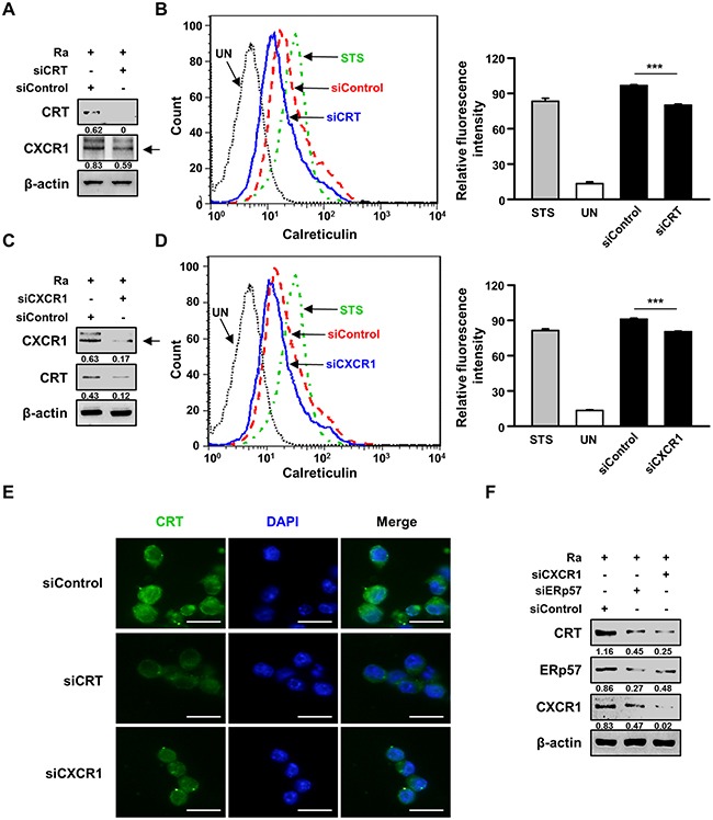
Figure 4. The interaction between CRT and CXCR1 activates the extrinsic apoptotic pathway during Mtb infection.

(A-E) Raw 264.7 cells were transfected with an siRNA targeting CRT (siCRT) or CXCR1 (siCXCR1) or with a non-specific siRNA (siControl) and then infected with Mtb H37Ra (MOI=10) for 24 h. (A, C) The production levels of CRT and CXCR1 were then analyzed by western blot analysis. (B, D) The cell-surface expression of CRT was analyzed by flow cytometry. Staurosporine (STS; 500 nM, 18 h) served as the positive control. (E) CRT staining (green) was visualized by fluorescence microscopy. Cell nuclei were visualized by DAPI staining (blue). Scale bar: 20 μm. (F) Raw 264.7 cells were transfected with siCXCR1 or an siRNA targeting ERp57 (siERp57) and then infected with Mtb H37Ra (MOI=10) for 24 h. The production levels of CRT, ERp57, and CXCR1 were then analyzed by western blotting. Western blot data presented are representative of three independent experiments. Numbers below the blot indicate the intensities ratios of each target protein to the β-actin control in each lane. The data are means ± SD of three independent experiments. *p<0.05, **p<0.01, and ***p<0.001.
