
Figure 9.
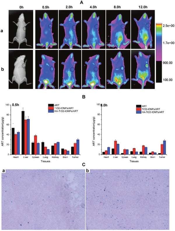
(A) In vivo NIR imaging of tumor-bearing mice intravenous injected with (a) free IR783 solution and (b) IR783-loaded HA-TiO2-IONPs at 0.5, 2, 4, 8 and 12h post injection; (B) Tissue distribution of ART, TiO2-IONPs/ART and HA-TiO2-IONPs/ART at 0.5h and 8h post injection; (C) The prussian blue staining images of (a) tumor tissue and (b) liver tissue for HA-TiO2-IONPs/ART group at 4h.