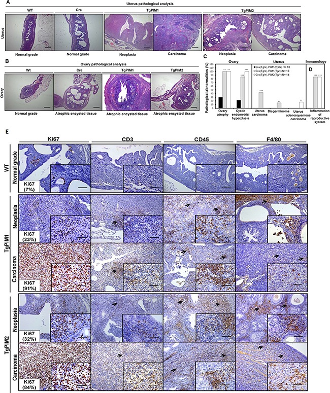
Figure 3. Pathological analysis of the uterus and ovary in TgPIM1/TgPIM2 transgenic models.

(A) Histopathological analysis of the uterus of WT and PIM1 and PIM2 transgenic females. Pictures depict representative observations in the different models. (B) Histopathological analysis of the ovary of WT, Cre and PIM1 and PIM2 transgenic females. Pictures illustrate representative observations in the different models. (C) Percentage of mice with pathological abnormalities observed. In each case, the statistics indicate the significance of the findings, compared to the WT females using Student's T test analysis, (*p < 0,05), (**p < 0,01) and (***p < 0,001). (D) Percentage of mice carrying inflammation in reproductive tissues. (E) Immunohistological characterization of immune infiltration in the uterus from MMTV-Cre/PIM1 and MMTV-Cre/PIM2 transgenic models. Pathological analysis of the uterus was performed by using both proliferation markers (Ki67) and immune mouse markers in order to determine whether the infiltration is lymphoid (CD3 and CD45) or macrophage (F4/80) origin. All markers are shown in WT, and both neoplasia and carcinoma stages for both transgenic models. The figure shows representative images of immune infiltration areas (marked with arrows in each marker). Scale bars indicate 50 μm (40×) in small-corned pictures and 100 μm (20×) in large pictures. All of the images were captured using the Microscope Olympus BX-61.
