
Figure 1. Immunophenotypic analysis of BMSCs and the construction of NF-200 promoter of lentivirus plasmid and identification.
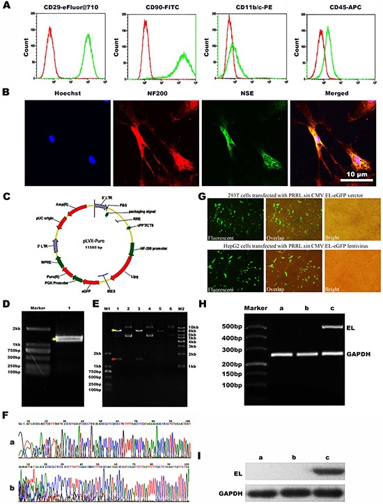
(A) Flow cytometry showed negative expression of CD45 and CD11b/c while positive expression of CD29 and CD90. (B) Confocal microscopy images to confirm the neurogenic differentiation of BMSCs by the neurocyte-specific markers NSE and NF-200. Hoechst33342 a blue tint to the nucleus, NF-200 dyed red, and NSE dyed green. (C) Scheme of plasmid. (D) Agarose gel electrophoresis of EL (endothelial lipase) DNA. (E) PRRL. sin. CMV. EL-eGFP digested with DraI and NotI enzymes1, 3, 5: DNA extracted from three stochastic monoclonal colonies of PRRL. sin. CMV. EL-eGFP; 2, 4, 6: Double digestion results of 1, 3, 5 with SpeI and EcoRI. (F) Sequencing of the recombined PRRL. sin. CMV. EL-eGFP verctor. a: The sequenceing after Pcmv; b: The sequenceing before IRES. (G) Production of recombined lentivirus and infection with HepG2 cells. a: Fluorescent image of 293T cells transfected with PRRL. sin. CMV. EL-eGFP verctor; b: Overlap image of 293T cells transfected with PRRL. sin. CMV. EL-eGFP verctor; c: Bright image of 293T cells transfected with PRRL. sin. CMV. EL-eGFP verctor; d: Fluorescent image of HepG2 cells transfected with PRRL. sin. CMV. EL-eGFP lentivirus; e: Overlap image of HepG2 cells infected with PRRL. sin. CMV. EL-eGFP lentivirus; f: Bright image of HepG2 cells transfected with PRRL. sin. CMV. EL-eGFP lentivirus. (H) Expressing of EL mRNA in HepG2 cells. a: untransfected HepG2 cells; b: transfected HepG2 cells with PRRL. sin. CMV. eGFP lentivirus; c: transfected HepG2 cells with PRRL. sin. CMV. EL-eGFP lentivirus; EL: endothelial lipase. (I) Expressing of EL protein in HepG2 cells. a. untransfected HepG2 cells; b. transfected HepG2 cells with PRRL. sin. CMV. eGFP lentivirus; c. infected HepG2 cells with PRRL. sin. CMV. EL-eGFP lentivirus.