
Figure 5. Immunofluorescence of spinal cord tissue.
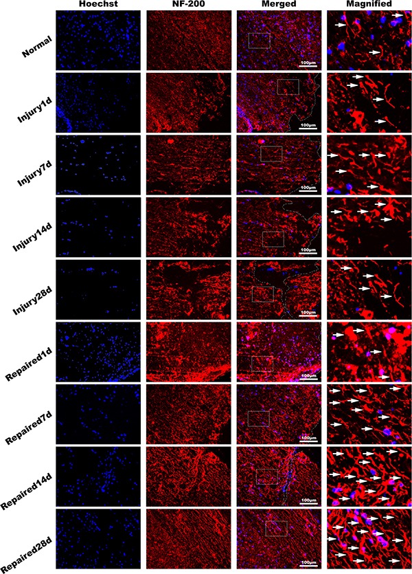
NF-200 antibody (red), dyeing Hoechst33342 nucleus (blue). It showed that the pathological changes in Injury group decreased with time obviously in longitudinal section. The NF-200 positive cells expressing quantity increased significantly in Repaired group compared to Injury group (n = 5). Bar: 100 μm.