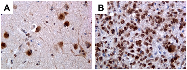
Figure 1. Immunohistochemical detection of PATZ1 in human GBM and perilesional normal cortex.

(A) Perilesional normal cortex: only neurons stain positively, mainly in the nucleus, while glial cells are negative. (B) Representative GBM: most of neoplastic cells are positive. Original magnification: 40x.
