
Figure 5. σ-R ligand PB28 attenuate mouse ERG possibly through Kv2.1 inhibition.
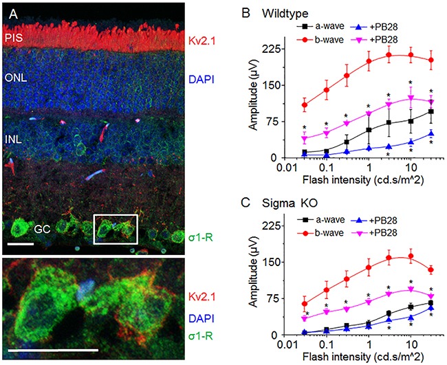
(A) Micrograph of mouse retina showing localization of Kv2.1 (red) and σ1-R (green). Nuclear layer is shown as blue DAPI staining. In the lower panel two enlarged ganglion cells marked with a white box showing Kv2.1 (red) and σ1-R (green) immunostaining. Scale bar 15 μM. (B) Average response of a- and b-wave amplitude in relation to light flash intensity. Vehicle injected eye response is shown as black (a-wave) and red (b-wave) traces. PB28 injected eye is shown as blue (a-wave) and purple (b-wave) traces. (C) Average ERG a- and b-wave responses from σ1-R knock out mice after saline or PB28 injection. Color representation as in B. Data is represented as mean ± SEM from at least 5 observations for each point and *P < 0.05 defines significance compared to control.