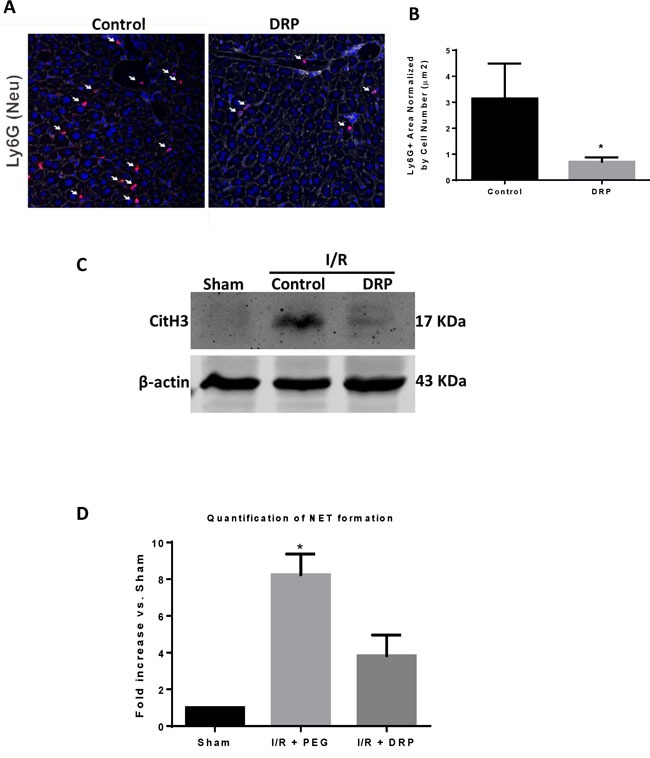
Figure 2. DRPs decrease neutrophil infiltration and neutrophil extracellular trap formation after I/R.

A. and B. Using confocal microscopy, there is a significant decrease in infiltrating neutrophils 6 hours after mice were subjected to I/R in DRP treated mice compared to mice that received control (mean 0.8 μm2 Ly6G+ area/total cells versus 3.1 μm2 Ly6G+ area/total cells, p < 0.001). Ly6G (red), nuclei (blue). Scale Bars 100μm. C. Cit-H3 protein levels were determined by Western blot in sham, I/R + control PEG, and I/R + DRP mice groups 6 hours after liver I/R. D. NETs acutely form in liver tissue 6 hours after liver I/R as assessed by serum levels of MPO-DNA. Treatment with DRPs after liver I/R resulted in a significant decrease in the levels of serum MPO-DNA at 6 hours. Results are expressed as the relative folds increase of MPO-DNA levels compared with sham; mean±SEM (n = 6/group). *P < 0.05.
