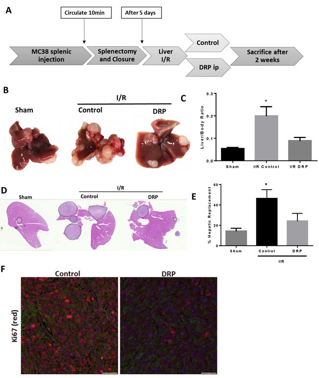
Figure 5. DRPs halt the I/R-induced accelerated growth of established liver micrometastases.

A. Schematic representation of the experimental design is depicted. Intrasplenic injection of MC38 cells was performed and metastatic tumor was allowed to grow for 5 days before the mice were subjected to liver I/R. DRPS or control PEG were given at the time of liver I/R. At 3 weeks, the mice were sacrificed and tumor growth was assessed. B. Representative images of hepatic nodules after necropsy in mice subjected to sham or I/R with or without DRP treatment. C. Liver I/R resulted in a significant increase in tumor growth at three weeks compared to sham mice seen by liver-to-body ratio. Treatment with DRPs after I/R resulted in a significant decrease in growth of already established micrometastases. D. and E. DRP treatment resulted in a significant decrease in tumor burden at three weeks as seen by percentage hepatic replacement by metastatic tumor. F. Treatment with DRPs during I/R significantly attenuated tumor cell proliferation three weeks later as seen by decreased Ki67 staining (mean 2.62 Ki67+ nuclei/ Area Actin 105μm2 I/R control versus 8.12 Ki67+ nuclei/105μm2 in I/R DRP group, p < 0.001). Ki67 (red), nuclei (blue), actin (green). Scale Bars 100μm.
