Figure 3.
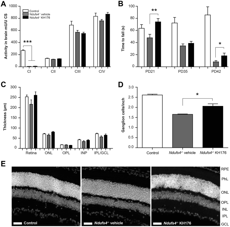
Improved motor function and reduced degeneration of ganglion cells in Ndufs4 −/− mice. (A) Activities of the separate respiratory chain complexes were measured in whole brain homogenates of control (white bar), Ndufs4 −/− vehicle-treated (gray bar) and KH176-treated (black bar) mice (at PD > 42). As expected, complex I was almost absent in brain tissue of Ndufs4 −/− mice, however, detectable. Activity (mU/U CS) as mean ± SEM (n = 8). Multivariate ANOVA (***p < 0.001). (B) Rotarod performance of control (white bar), Ndufs4 −/− vehicle-treated (gray bar) and KH176-treated (black bar) mice at PD21, PD35 and PD42. KH176 treatment significantly improved motor performance in Ndufs4 −/− mice on PD21 and PD42. Time to fall (s) expressed as mean ± SEM (n = 15). Multivariate ANOVA (**p < 0.01, *p < 0.05). (C) Thickness of complete retina and its different layers; outer nuclear layer (ONL), outer plexiform layer (OPL), inner nuclear layer (INL) or the combined inner plexiform layer with the ganglion cell layer (IPL/GCL). Control (white bar), Ndufs4 −/− vehicle-treated (gray bar) and KH176-treated (black bar) mice (at PD > 42). Thickness (µm) expressed as mean ± SEM (n = 3–4). (D) Number of retinal ganglion cells expressed as mean ± SEM (n = 3–4). Control (white bar), Ndufs4 −/− vehicle-treated (gray bar) and KH176-treated (black bar) mice (at PD > 42). Kruskal-Wallis (*p < 0.05). (E) Representative images of the retina (scale: 50 µm). Different layers: retinal pigment epithelium (RPE), photoreceptor layer (PhL), outer nuclear layer (ONL), outer plexiform layer (OPL), inner nuclear layer (INL), inner plexiform layer (IPL) and ganglion cell layer (GCL).

