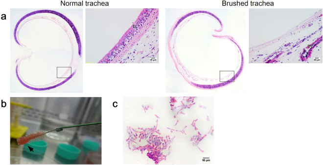
Figure 1.
(a) H&E stained trachea sections from the normal and injury groups for direct and indirect co-culture assays: (left panel) Normal group with intact pseudostratified epithelium, and (right panel) injury group following brushing-induced tracheal injury. Insets show the epithelium layer at higher magnification. (b) Brushing technique removes tracheal epithelium layer. (c) H&E stained cell collection from tracheal brushing, which predominantly consists of ciliated cells (scale bars (a) = 20 μm, (c) = 50 μm).

