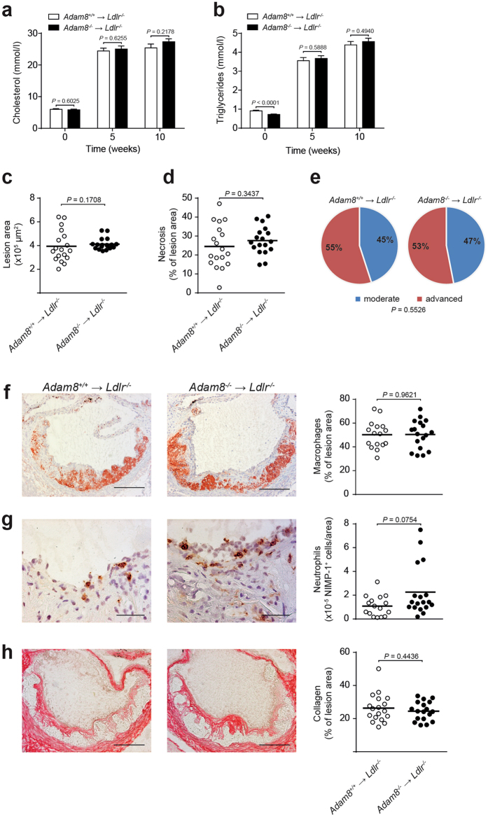
Figure 3.
Hematopoietic ADAM8 deficiency in Ldlr −/− mice does not affect size or morphology of advanced atherosclerotic lesions. (a,b) Plasma cholesterol (a) and triglyceride (b) levels after 0, 5 and 10 weeks of western type diet (WTD) feeding in female Ldlr −/− chimeras with (Adam8 −/− → Ldlr −/−) or without (Adam8 +/+ → Ldlr −/−) hematopoietic ADAM8 deficiency (n = 20 mice per genotype, parametric Student’s t-test). (c, d) Quantification of the aortic root lesion area (c, non-parametric Mann-Whitney U test) and necrotic core area (d, parametric Student’s t-test) of Adam8 +/+ → Ldlr −/− and Adam8 −/− → Ldlr −/− mice (n = 18 mice per genotype) after 10 weeks of WTD. (e) Plaque progression stage (n = 51 aortic root atherosclerotic lesions per genotype) was scored (Fisher’s exact test). (f–h) Representative examples of (immuno)histochemical stainings and quantifications for MOMA-2+ macrophages (f, n = 18/16 mice, parametric Student’s t-test, scale bar, 200 μm), NIMP+ neutrophils (g, n = 18/16 mice, non-parametric Mann-Whitney U test, scale bar, 50 μm) and Sirius Red stained collagen (h, n = 18/16 mice, parametric Student’s t-test, scale bar, 200 μm) in the aortic root after 10 weeks of WTD feeding.

