. 2017 Sep 15;6:e29236. doi: 10.7554/eLife.29236

Figure 7. Intracellular localization of NSs in Vero-E6 cells infected with rMP12 or rMP12 variants encoding NSs variants with interface residue mutations (muT1, muT3, muT4).

Cells were fixed 24 h p.i. and co-stained with either anti-N or anti-NSs antibodies (green) as indicated and DAPI (blue).

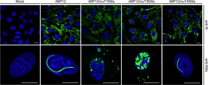
Figure 7.

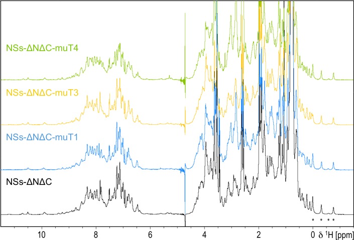
Figure 7—figure supplement 1. 1D 1 hr NMR spectra of NSs-ΔNΔC and interface mutation variants.

Figure 7—figure supplement 1.

Asterisks indicate distinct high-field shifted methyl resonances with conserved chemical shifts in all variants.