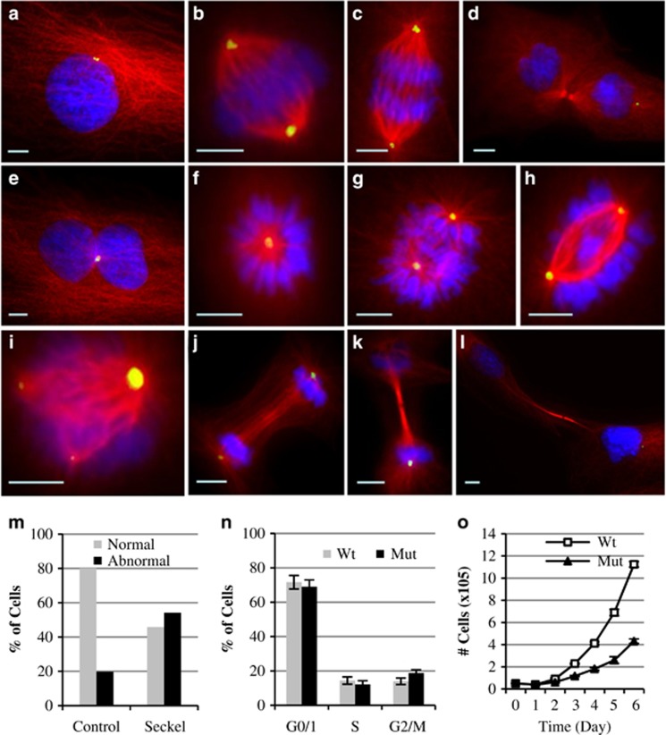
Figure 3.
Mitotic morphology, cell cycle and doubling time analyses of PLK4-Seckel fibroblast cells. Immunostaining of PLK4-Seckel cells with α-tubulin (red) and pericentrin (green) antibodies. Nuclei were counterstained with DAPI (blue). (a–d) Morphologies of interphase, metaphase, anaphase and telophase of control fibroblasts cells, respectively. PLK4-Seckel cells; (e) with two nuclei and one centrosome in interphase, (f) monopolar spindle in metaphase, (g–h) uneven aligned bipolar spindle with improperly aligned chromosomes in metaphases, (i) tripolar mitotic spindle with differently sized centrosomes, (j) delayed cleavage furrow with irregular spindle alignment in telophase, (k–l) extended telophase bridge with midbody ring in telophase. Scale bars, 5 μm. (m) Percentage of cells showing abnormal mitotic morphology in PLK4-Seckel fibroblasts. (n) Increased G2/M phase in PLK4-Seckel cells, P=0.006 (T-test), error bars represent SD. (o) Increased doubling time of PLK4-Seckel fibroblasts compared to control.

