
Fig. 1.
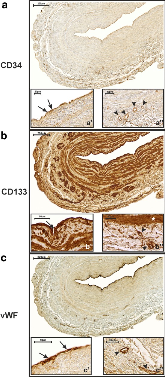
Immunohistochemical detection of CD34 (a), CD133 (b) and vWF (c) expression in a saphenous vein graft obtained from a 59-year-old CABG patient who developed a complete graft occlusion 11 months after surgery. CD34 is expressed within endothelial cells present in the tunica intima (a′ arrows) of the studied graft, as well as in small blood vessels and capillaries present in the tunica adventitia (a″ arrow heads). CD133 is strongly expressed in intimal endothelial cells (b′ arrow) and smooth muscle cells (b″ asterisk) (IRS = 12). Small adventitial blood vessels and capillaries are also CD133-positive (b″ arrow heads). Finally, vWF factor immunoreactivity is observed within intimal endothelial cells (c′ arrows). Only a very few small blood vessels, as are present in the adventitia, are vWF-positive (c″ arrow heads)