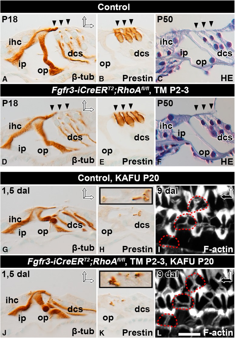
Figure 4.
RhoA is dispensable for supporting cell maturation and wound healing. Recombination was induced in auditory SCs of RhoAfl/fl;Fgfr3-iCre-ERT2 mice at P2–3, and analysis was performed at adulthood. Paraffin-embedded cross-sections (A–H, J, K) and phalloidin-labeled whole-mount specimens (I, L) of the organ of Corti of the cochlear medial turn of mutant and control mice. A, D, At P18, β-tubulin immunocytochemistry shows no major structural differences between SCs of the two genotypes. B, E, Prestin immunostaining shows comparable morphology of non-recombined OHCs as well. C, F, Hematoxylin staining shows that the cytoarchitecture of the organ of Corti is comparable between mutant and control mice at P50. G, J, Most OHCs are lost 1.5 d after induction of ototoxic lesion (KAFU, see Methods). In both types of specimens, β-tubulin–positive SCs maintain positions at the reticular lamina, although their cell bodies have partially lost upright position. H, K, In both genotypes, Deiters cells phagocytose prestin-positive OHC debris, shown in insets at a higher magnification. I, L, Both control and RhoA-depleted SCs form stable F-actin scars, shown 9 d postlesion. Red dashed lines mark scars at the site of lost OHCs. Abbreviations: β-tub, β-tubulin; dal, days after lesion; dcs, Deiters cells; HE, hematoxylin; ihc; inner hair cell; i.p., inner pillar cell; KAFU, kanamycin and furosemide; ohc, outer hair cell; op, outer pillar cell; TM, tamoxifen. Scale (in L): A–H, J, K, 20 µm; I, L, 6.5 µm.

