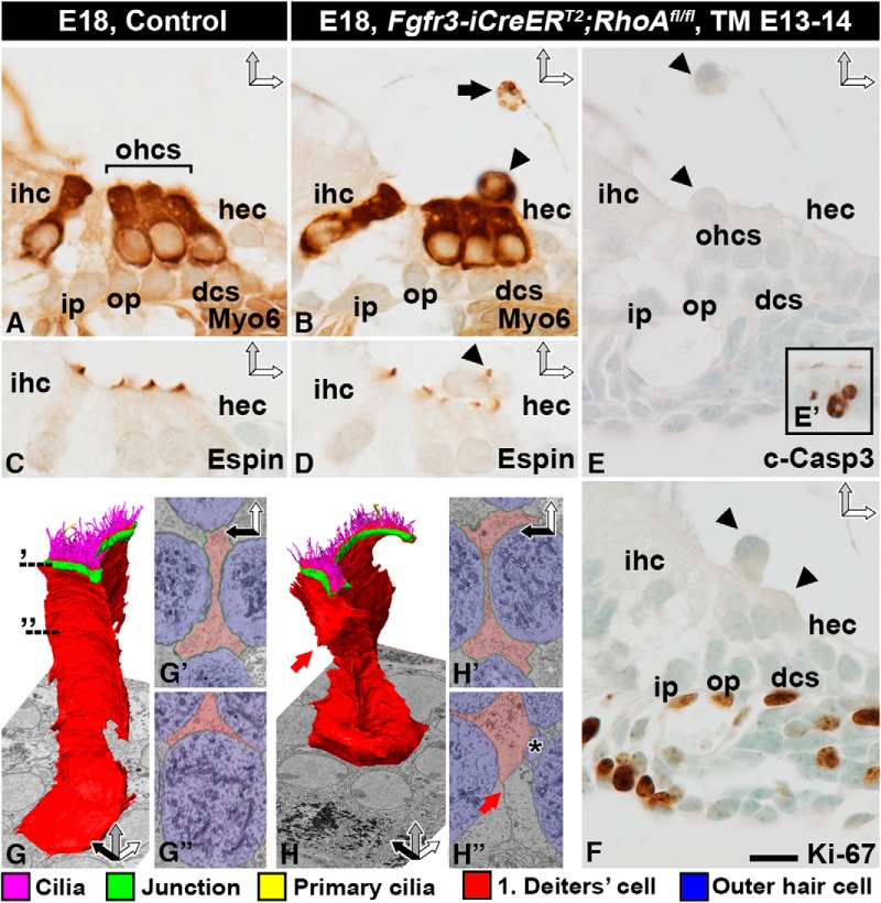
Figure 5.
RhoA-depleted outer hair cells are extruded from the organ of Corti. Recombination was induced in RhoAfl/fl;Fgfr3-iCre-ERT2 mice at E13–14, and analysis was performed at E18.5. Images show paraffin-embedded cross-sections from the medial turn of the cochlea (A–F) as well as single block-face images and 3D modeling of Deiters cells (G–H″). A, B, Myosin 6–immunostained OHCs extrude (arrowhead in B) from the organ of Corti of mutant mice. The presence of myosin 6–positive degenerating cell profiles (arrow in B) in the endolymph shows that OHCs die after extrusion. C, D, Similar to controls, OHCs of mutant cochleas possess espin-positive stereociliary bundles with abnormal morphology. Arrowhead points to an extruding OHC with an espin-stained bundle (D). E, E′, Extruding OHCs (arrowheads) are negative for cleaved caspase-3. As a positive control in the same section, the inset shows apoptotic cell profiles in the greater epithelial ridge of the cochlea. F, Extruding OHCs are negative for Ki-67. Proliferating mesenchymal cells underneath the basement membrane in the same section serve as positive controls. G–G″, 3D modeling of a control Deiters cell. Dashed lines demonstrate the level of block-face single images (G′, G″). H–H″, 3D modeling of a mutant Deiters cell shows that the general features of this cell type are maintained, but apical junctions (H′) and the basolateral cell membrane (red arrow in H, H″) have moved due to the loss of adjacent OHCs. A Deiters cell has invaded the space previously occupied by an OHC, and only a “tail” (asterisk) is left of the extruding OHC (H″). Color coding is explained below SBEM images. Abbreviations: c-Casp3, cleaved caspase-3; dcs, Deiters cells; hec, Hensen cell; ihc; inner hair cell; i.p., inner pillar cell; Myo6, myosin 6; ohc, outer hair cell; op, outer pillar cell; TM, tamoxifen. Scale bar (in F): A–F, 10 µm; G, H, 4 µm; G′, G″, H′, H″, 3 µm.

