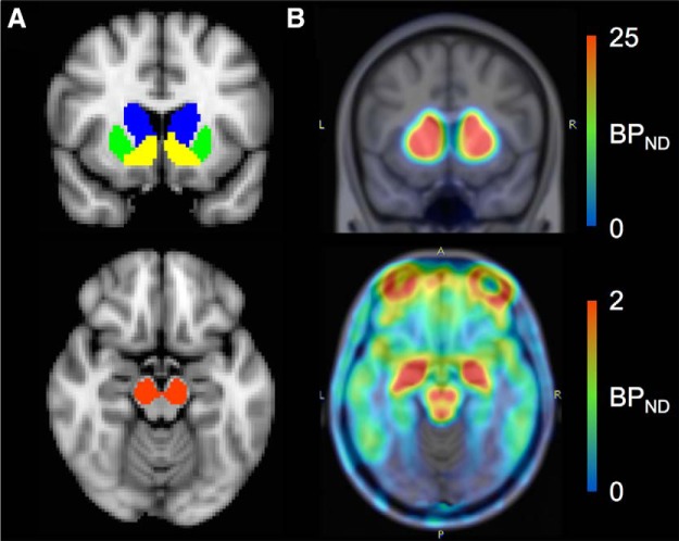
Figure 1.
[18F]fallypride BPND images reflecting DRD2 availability. A, Shown are ROIs from which mean BPND were extracted for analyses: caudate (blue), putamen (green), ventral striatum (yellow), and midbrain (red). B, Example of a [18F]fallypride BPND image showing high BPND in the striatum (top) and midbrain (bottom).

