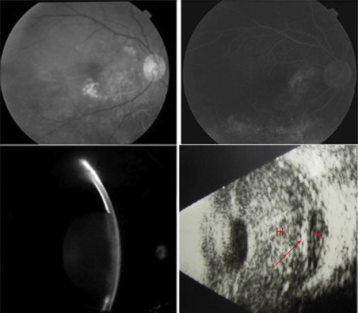
Fig. 1.

Fundus photography of the right eye showing numerous small-sized and confluent drusen (upper left), which were stained at the late stage without leakage using fluorescein angiography (upper right). These pictures were obtained 2 years prior to the episode. Slit-lamp examination shows forward displacement of the iris and lens with total iridocorneal touch (lower left). B-Scan ultrasonography of the right eye reveals a dome-shaped choroidal detachment (arrow) with hypo-echogenous content (asterisk) in addition to a dense vitreous hemorrhage (H) (lower right).
