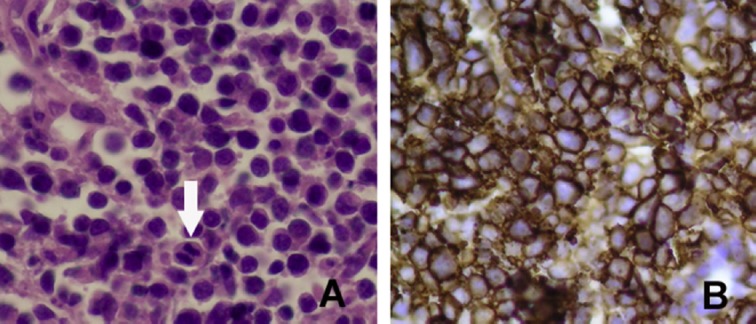
Fig. 3.

(A) Histopathological studies reveal diffuse infiltration of medium-sized to large neoplastic cells with high mitotic activity (arrow; hematoxylin and eosin stain, ×400). (B) The immunohistochemical stain shows the neoplastic cells with CD20 (+) (×400).
