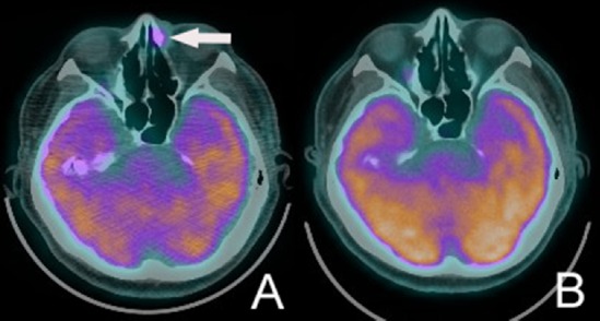
Fig. 4.

(A) Whole-body fludeoxyglucose positron emission tomography (FDG PET) scan demonstrates focal increased FDG uptake in the inferior medial aspect of left orbital cavity (arrow). (B) One year after rituximab, cyclophosphamide, doxorubicin, vincristine, and prednisone (R-CHOP) therapy, no abnormal glucose metabolic region is found in the whole-body FDG PET scan.
