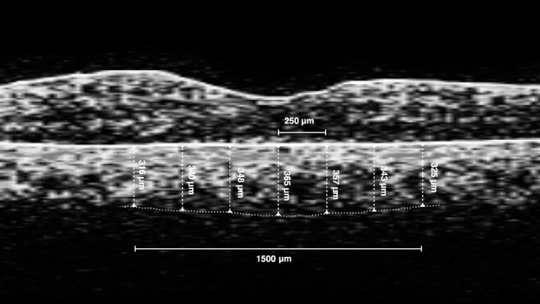
Fig. 2.

Example of measurement of subfoveal choroidal thickness. A one-line raster scan optical coherence tomography image was taken through the foveal center horizontally. The subfoveal choroid was defined as the choroid beneath the concave central retinal depression 1500 μm in diameter. A single choroidal thickness measurement was obtained for the horizontal raster from the outer border of the retinal pigment epithelium to the inner scleral border. The manual caliper was used to sequentially measure 250-μm distances in both radial directions (total of 7 measurements/patient) by two independent observers. The average of these measurements was calculated as subfoveal choroidal thickness. As shown in this patient, the subfoveal choroidal thickness was 342 μm.
