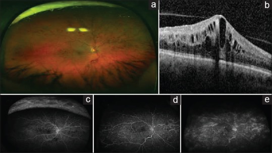
Figure 1.

Retinal imaging of central retinal vein occlusion. (a) The ultra-wide fundus photograph shows numerous intraretinal hemorrhages and vascular tortuosity consistent with central retinal vein occlusion. (b) The ocular coherence tomography scan shows intraretinal edema with thickening in the central macula. (c-e) The fluorescein angiogram progresses with time from left to right. The fluorescein angiogram shows leakage to the macula, perivascular leakage in the periphery, and shunt vessels due to old central retinal vein occlusion
