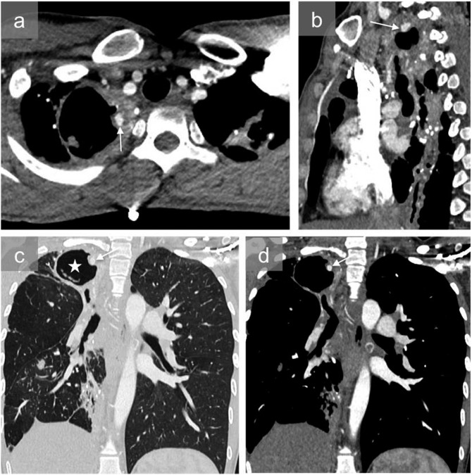
Figure 8.
A 45-year-old male with a history of pulmonary tuberculosis and recurrent tuberculosis under antibiotics who was admitted to the hospital for acute haemoptysis: axial (a), sagittal (b) and coronal mediastinal and pulmonary images at the same level (c, d) show a large right apical cavity (white star) and pseudoaneurysm in the wall of the cavity (arrows). The patient was successfully treated with coil embolization.

