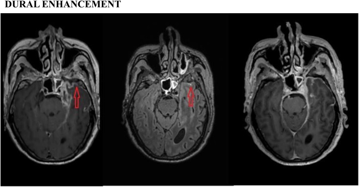
Figure 4.
Dural enhancement: post-contrast three-dimensional (3D)-T1 sampling perfection with application optimized contrast using different flip-angle evolutions (SPACE) (left), 3D-T2 fluid-attenuated inversion recovery (FLAIR) (middle) and 3D-T1 magnetization-prepared rapid gradient-echo (MPRAGE) (right) images showing abnormal dural enhancement (arrows) in the left temporal region in a patient with tubercular meningitis. However, the post-contrast T1-SPACE image is much more conspicuous than the post-contrast 3D-T1-MPRAGE and 3D-T2-FLAIR images.

