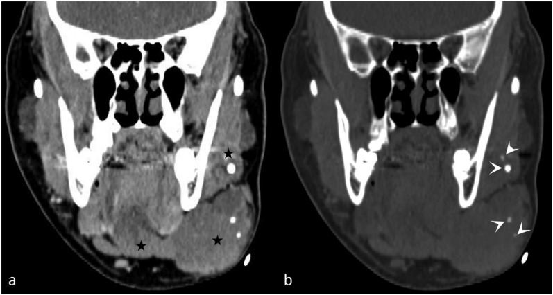
Figure 2.
A large facial venous vascular malformation with phleboliths: (a) the coronal contrast-enhanced CT image shows a soft-tissue mass (stars) infiltrating the left submandibular gland. (b) The coronal virtual non-contrast image reveals multiple phleboliths (arrowheads) in the mass, aiding in the diagnosis of a venous vascular malformation.

