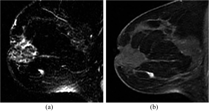
Figure 5.
A 43-year-old female with extensive ductal carcinoma in situ in the opposite breast; work-up for extent of disease. (a) T2 weighted image shows solitary lesion with high signal. (b) Irregular enhancing mass on T1 fat-suppressed first post-contrast image. Biopsy showed a 6-mm poorly differentiated invasive ductal carcinoma.

