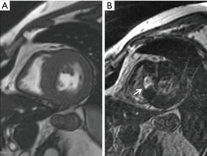
Figure 2.

Cardiac magnetic resonance. (A) Cine image in short axis view. Severe midventricular septal hypertrophy is seen; (B) at the same level, late enhancement imaging showing focal area in the right side of midventricular septum, with bright (almost transmural) enhancement secondary to induced infarction from the alcohol septal ablation (arrow).
