Figure 2.
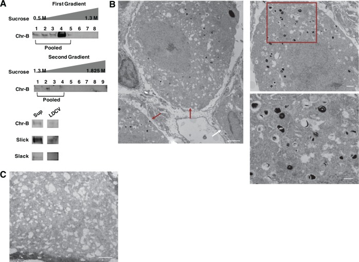
Slick channels are localized to Large Dense-Core Vesicles. (A) LDCV fractionation assay of adult mouse DRGs. Chromogranin-B (Chr-B) used as positive indicator for LDCV. Final pooled elution tested for Chr-B, Slick (anti-mouse), and Slack (anti-mouse) proteins compared to supernatant (Sup) by immunoblot. (B) Representative electron micrography (EM) of adult mouse lumbar DRGs labeled with Slick (anti-mouse) and enhanced with 3,3′-Diaminobenzidine (DAB). Left image indicates staining in small- and medium-sized cell bodies (red arrows), with a nonlabeled cell body (white arrow). Scale bar represents 2 μm (left and top right) and 500 nm (bottom right). Right image enhanced from center image (red box). (C) Representative image of negative control staining adult mouse lumbar DRG no primary with DAB staining. Scale Bar 2 μm.

