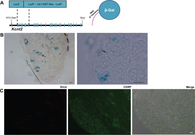
Figure 4.
Slick KO mice confirm Slick localization to neuronal subtype. (A) Illustration of Slick-LacZ global KO mice, exons 2 to 7 replaced with LacZ cassette. Right: representation of resultant β-galactosidase protein with partial Slick N-terminal. (B) Representative images of Slick KO mouse DRG X-Gal stain at low and higher magnification. Arrows indicate “punctate X-Gal” staining of the resultant β-galactosidase protein with partial Slick N-terminal. Scale bars: 20 μm. (C) Representative immunohistochemical images of Slick KO mouse DRG exhibiting no Slick (anti-mouse) immunoreactivity. Merge with DAPI and DIC. Scale bars: 20 μm. DAPI, 4,6-diamidino-2-phenylindole dihydrochloride; DIC, differential interference contrast; DRG, dorsal root ganglia; HO, knockout.

