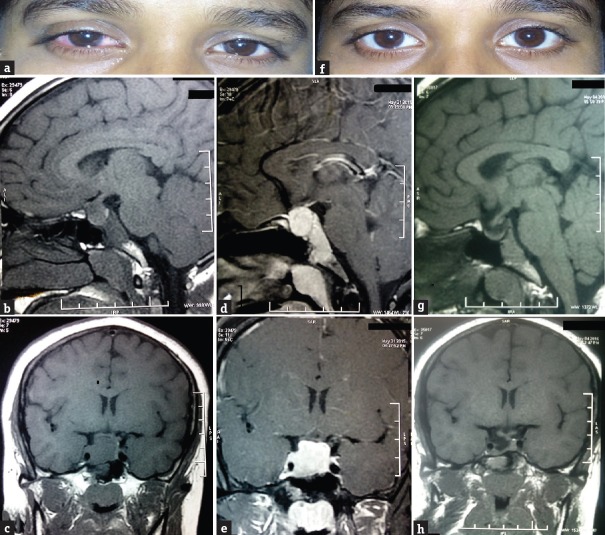
Figure 1.
(a) Lacrimation and redness of right eye during headache. (b) T1-weighted image showing a macroadenoma (1.8 cm × 1.7 cm × 2.3 cm). (c) T1-weighted image showing macroadenoma causing stalk compression and parasellar extension. (d) Postcontrast image showing intense enhancement of macroadenoma. (e) Postcontrast image showing intense enhancement of macroadenoma. (f) Recovery after drug treatment. (g) T1-weighted image showing a significant tumor size reduction after treatment. (h) T1-weighted image showing significant tumor size reduction after treatment

