. 2017 Aug 11;6:e27793. doi: 10.7554/eLife.27793

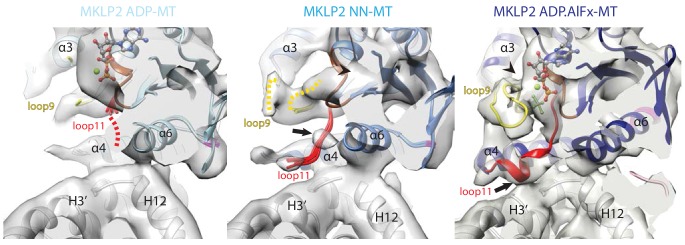
Figure 3. Conservation and divergence at the MKLP2-MD nucleotide binding pocket.

View of the MKLP2-MD nucleotide binding pocket showing nucleotide-dependent transitions in helix-α4, P-loop (brown), loop9 (yellow) and loop11 (red). In the ADP state (left), density corresponding to ADP is visible and connected to the P-loop, while in the NN state (middle), density for nucleotide is no longer present (arrowhead). In the ADP.AlFx state, a hydrolysis-competent ‘closed’ nucleotide pocket conformation is observed, loop11 becomes fully ordered with a single helical turn contacting α–tubulin H3’ (arrow), and loop9 forms an ordered β-hairpin which contacts the nucleotide (arrowhead).

Figure 3.

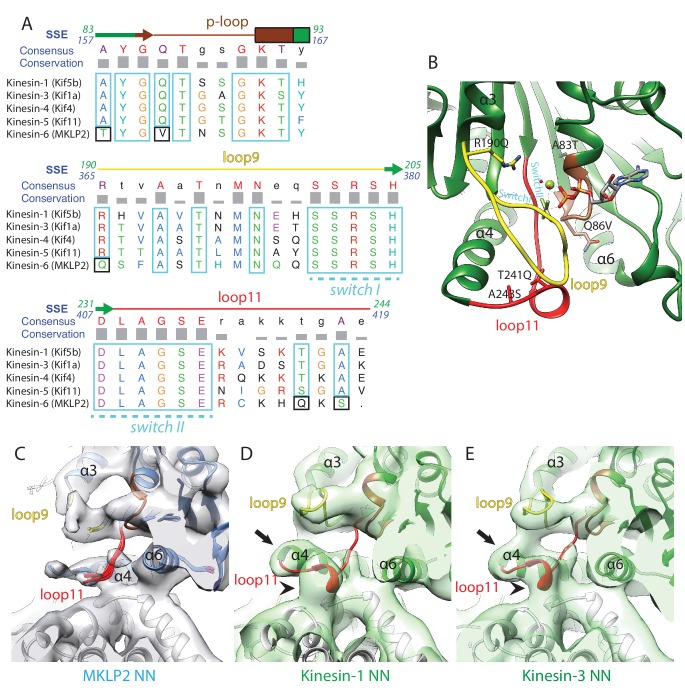
Figure 3—figure supplement 1. Comparison of MKLP2 nucleotide binding site with other N-kinesins.

Figure 3—figure supplement 1.

(A) Sequence alignment of important regions at the nucleotide pocket for kinesin-6 MKLP2 and representative members from other Kin1/3/4/5 for which X-ray structures are available. Residue letters are coloured according to their properties according to the Clustal X scheme (Larkin et al., 2007). Information on consensus, conservation and secondary structure is shown above the alignments. Sequence numberings for Kif5B (Kin1, green) and MKLP2 (kinesin-6, blue) are shown adjacent to the secondary structure schematics. Highly conserved/similar kinesin residues are boxed in light blue, whereas highly conserved kinesin residues which have diverged are boxed in black. The signature kinesin switch I and II sequences are indicated. (B) The nucleotide pocket of the X-ray structure of tubulin-bound Kin1 in the presence of ADP.AlFx (PDB 4HNA, [Gigant et al., 2013]). The position of the switch I/II sequences are annotated in cyan, whilst those that have diverged in MKLP2 are shown in sticks and annotated in black, in format Kin1 amino-acid, Kin1 amino-acid number, mouse MKLP2 amino-acid. (C–E) The nucleotide binding regions in the NN states of (C) kinesin-6 MKLP2, (D) Kin1 Kif5a and (E) Kin3 Kif1A (Atherton et al., 2014) are shown in their corresponding experimental densities (grey density for MKLP2-MD, green densities for Kin1 and Kin3). Arrows indicate full extension of helix-α4’s N-terminus in this state and arrowheads indicate formation of a single-turn helix in loop11 for Kin3 and Kin1 that contacts the MT, in contrast to MKLP2 where these regions are less ordered.
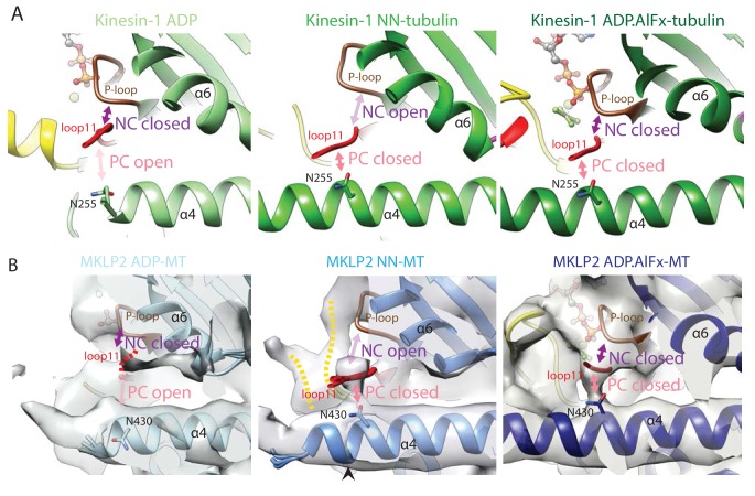
Figure 3—figure supplement 2. Conservation of nucleotide-dependent subdomain movements and location and configuration of the clefts that separate them in Kin1 and MKLP2.

Figure 3—figure supplement 2.

(A). The NC and PC in Kin1 ADP (PDB: 1BG2), NN (PDB: 4LNU) and ADP.AlFx crystal structures (PDB: 4HNA). With ADP, the NC is closed (loop11 contacts the P-loop) while the PC is open (loop11 does not contact helix-α4). In the NN state, the NC opens (loop11 releases its contact with the P-loop and instead associates with helix-α4) and the PC closes. In the ADP.AlFx state the PC remains closed and the NC closes as loop11 contacts both the P-loop and helix-α4. The position of N255, which reorientates upon nucleotide release to contact loop11 and close the polymer cleft is shown (Shang et al., 2014). (B) MKLP2 follows the same state-dependent sequence of changes at the NC and PC clefts as Kin1. MKLP2-MD cryo-EM density is shown in transparent grey with the same views shown as for Kin1 in panel A. The predicted position of N430 (corresponding to kinesin-1’s N255) in the models is indicated.
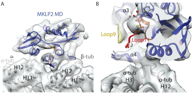
Figure 3—figure supplement 3. MT-bound MKLP2-MD-AMPPNP does not adopt an ‘ATP-like state’.

Figure 3—figure supplement 3.

(A) Side profile of the asymmetric unit of the MKLP2-MD-AMPPNP reconstruction, showing MT binding site similar to that seen in other nucleotide states. The fitted MKLP2-MD-AMPPNP model is in blue, the α-tubulin model is in light grey and the β-tubulin model is in dark grey. (B) View, similar to Figure 3C, of the nucleotide-binding site of the MKLP2-MD-AMPPNP reconstruction.