. 2017 Aug 11;6:e27793. doi: 10.7554/eLife.27793

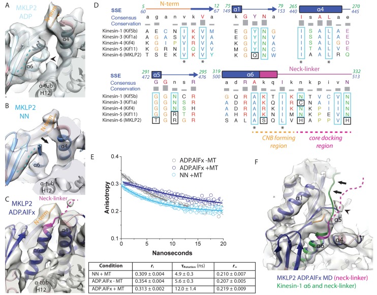
Figure 6. MKLP2 neck-linker response to nucleotide.

(A) MKLP2-MD-ADP has a short helix-α6 leaving a gap between its terminus and helix-α4 (arrow), showing the cleft for the neck linker is closed and the CNB is not formed. Helix-α6 has relatively poor density, suggesting it may be partially disordered at its C-terminus (arrowhead). Density that could flexibly accommodate part of the neck linker is present connected to β-sheet1 (blue dotted line). (B) MKLP2-MD-NN also has a short helix-α6 leaving a gap between its terminus and helix-α4 (arrow). (C) MKLP2-MD-ADP.AlFx has an extended helix-α6, the initial portion of the neck-linker (magenta) inserts between the N-terminus (orange) and helix-α4 forming the CNB (arrowhead). The remaining portion of the neck-linker is flexible. (D) Sequence alignments of representative members (in brackets) of the Kin1/3/4/5/6 families for the neck-linker and its contact regions. Residue colouring using the Clustal X scheme (Larkin et al., 2007). Sequence numberings for Kif5b (Kin1, green) and MKLP2 (kinesin-6, blue) are shown adjacent to the secondary structure schematics. Well conserved residues in MKLP2 are boxed in light blue, whereas otherwise well conserved residues which have diverged in MKLP2 are boxed in black (see also Figure 6—figure supplement 2). Asterisks indicate conserved hydrophobic residues participating in CNB formation. Regions of the neck-linker involved in CNB formation or core docking are indicated by dashed lines in orange and magenta, respectively. (E) Time-resolved fluorescence anisotropy of FlAsH labeled MKLP2-MD: (1) NN + MT, light blue; (2) ADP.AlFx –MT, grey and (3) ADP.AlFx +MT, dark blue. Data shown in the table below are representative of 5 replicate samples. (F) MKLP2-MD-ADP.AlFx neck-linker in magenta (disordered region, dashed line). Kin1 helix-α6 and neck-linker (dark green) has been superimposed on helix-α6 of MKLP2-MD, showing the expected position of the neck-linker. There is no density corresponding to a docked neck-linker (arrows) suggesting it is mainly disordered. A small amount of density close to helix-α5 (arrowhead) likely indicates that alternative conformations are flexibly explored.

Figure 6.

Figure 6—figure supplement 1. The neck-linker in MT-bound MKLP2-MD-AMPPNP is not directed towards the MT plus end.
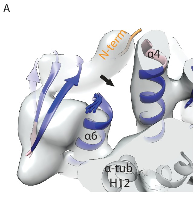
Figure 6—figure supplement 1.

(A) View, similar to Figure 6A–C, of the N-terminus, helix-α6 and neck-linker of the MKLP2-MD-AMPPNP reconstruction. The arrow indicates a gap between helix-α6 and helix-α4, suggesting a shortened helix-α6, a closed docking cleft and therefore no CNB formation or neck-linker docking. The fitted MKLP2-MD-AMPPNP model is in blue and the α-tubulin model is in light grey.
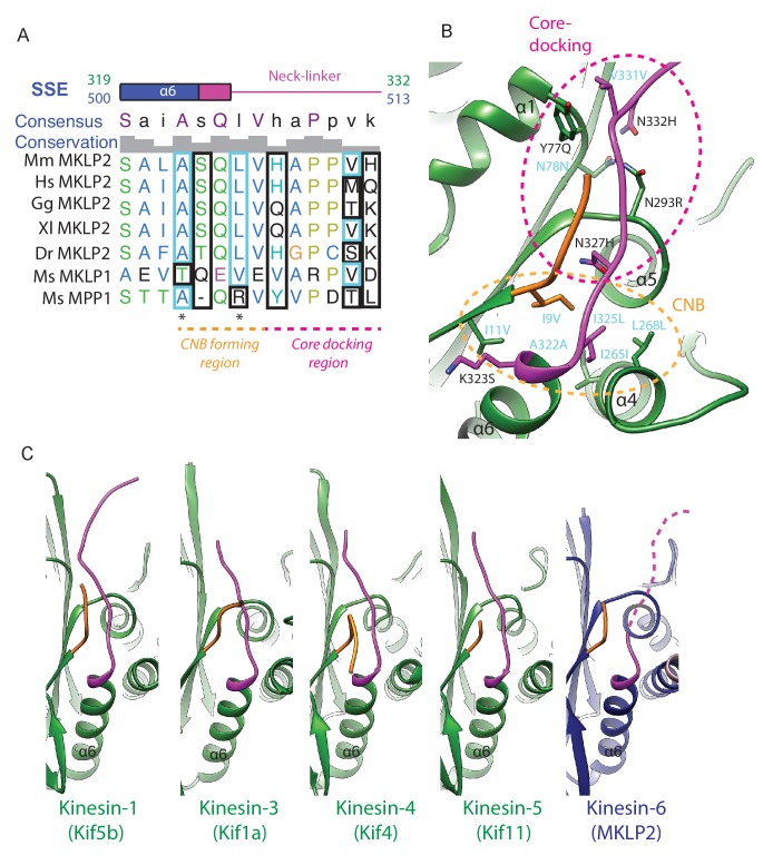
Figure 6—figure supplement 2. MKLP2’s neck-linker has an atypical sequence across species that precludes core-docking.

Figure 6—figure supplement 2.

(A) Sequence alignment of the mouse MKLP2 neck-linker sequence with the two other mouse kinesin-6s MKLP1 and MPP1 and MKLP2 from selected animal species. Species abbreviations are; Mm, Mus musculus, Hs, Homo sapiens, Gg, Gallus gallus, Xl, Xenopus laevis, Dr, Danio rerio. Residue letters are coloured according to their properties according to the Clustal X scheme (Larkin et al., 2007). Information on consensus, conservation and secondary structure is shown above the alignments. Sequence numberings for Kif5B (Kin1, green) and mouse MKLP2 (kinesin-6, blue) are shown adjacent to the secondary structure schematics. Highly conserved residues in plus end kinesins which remain conserved or have similar properties in kinesin-6s are boxed in light blue, whereas highly conserved kinesin residues which have diverged are boxed in black (see also Figure 6D). Conserved hydrophobic residues participating in forming the CNB are marked by asterisks. Regions of the neck-linker involved in CNB formation or core-docking are indicated by dashed lines in orange and magenta, respectively. (B) The cover-neck bundle (CNB) and docked-neck linker in the x-ray structure of tubulin-bound Kin1 Kif5b (PDB: 4HNA). Side chains are shown for conserved kinesin residues involved in CNB formation (region within the orange-dashed circle) and neck-linker docking (region within the magenta-dashed circle). Highly conserved residues that remain conserved or highly similar are annotated in cyan, whilst those that have diverged are annotated in black, in format Kin1 amino-acid, Kin1 amino-acid number, mouse MKLP2 amino-acid. (C) Full neck-linker docking is observed in all structures with an ATP analogue and a ‘closed’ hydrolysis-competent nucleotide pocket apart from kinesin-6 MKLP2; Kin1 Kif5b (PDB: 4HNA, [Gigant et al., 2013]), Kin3 Kif1a (PDB: 4UXR, [Atherton et al., 2014]), Kin4 Kif4 (PDB: 3ZFD, [Chang et al., 2013]), Kin5 Kif11 (PDB: 3HQD, [Parke et al., 2010]). For MKLP2, a dashed magenta line is used to represent neck-linker flexibility.