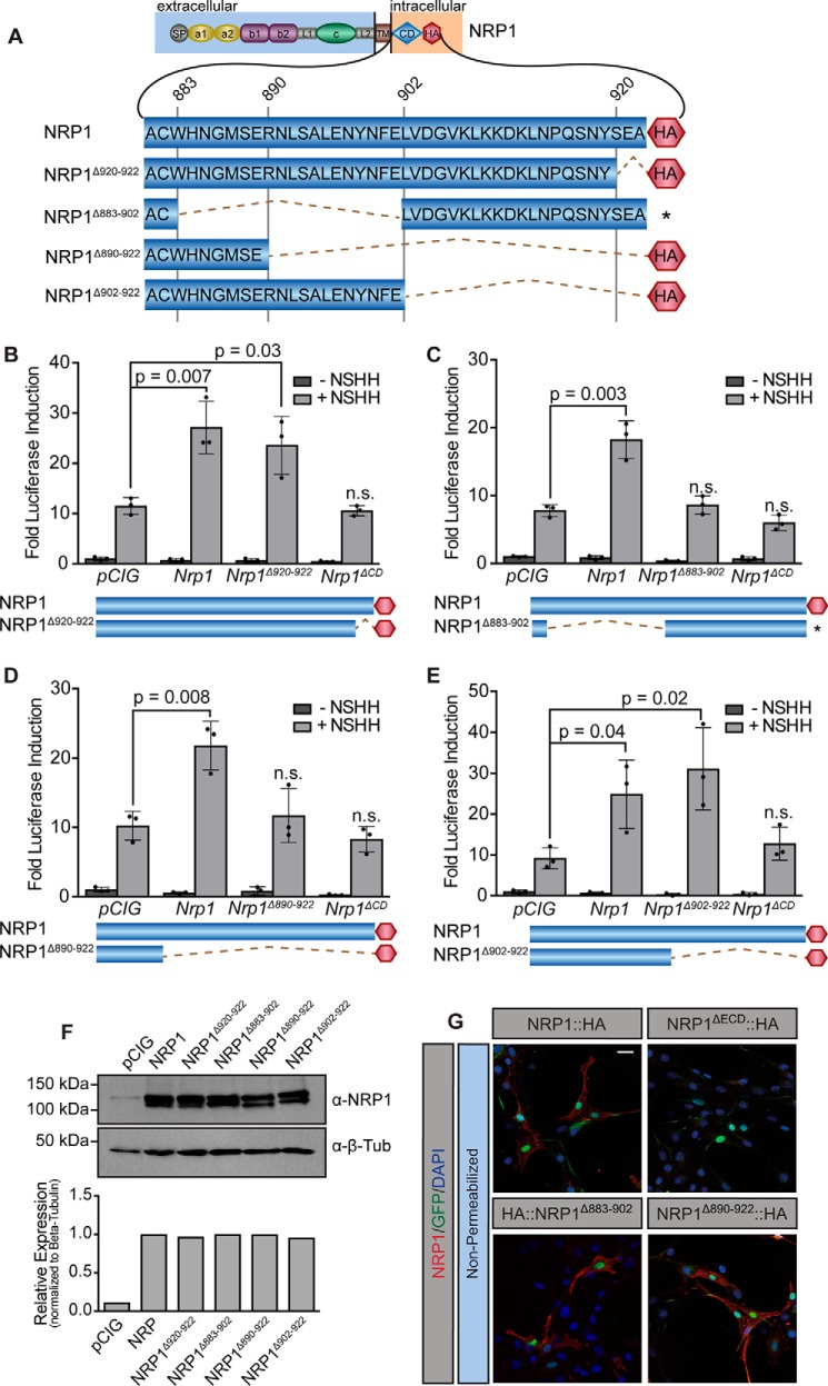
Figure 5.
Identification of a 12-amino acid motif in the NRP1 CD required for HH signal promotion. A, diagram of the NRP cytoplasmic domain, with amino acid number indicated (top) and deletions indicated by dotted lines. B–E, luciferase reporter activity in NIH-3T3 cells transfected with NRP constructs as indicated and stimulated with NSHH-conditioned medium (+NSHH). Data are reported as mean -fold induction ± S.D., with p values calculated using two-tailed Student's t tests. F, top, Western blot analysis of HA-tagged protein levels in NIH-3T3 cell lysates with detection of β-tubulin (β-Tub) as a loading control. Bottom, quantitation of NRP levels relative to β-tubulin. G, antibody detection of an extracellular NRP1 epitope (α-NRP1, red) in non-permeabilized NIH-3T3 cells to assess cell surface localization of NRP1, NRP1ΔECD, NRP1Δ883–902, and NRP1Δ890–922. Nuclear GFP (green) indicates transfected cells, whereas DAPI (blue) marks all nuclei. Note that NRP1ΔECD lacks the NRP1 antibody epitope and served as a negative control. Scale bar = 10 μm.

