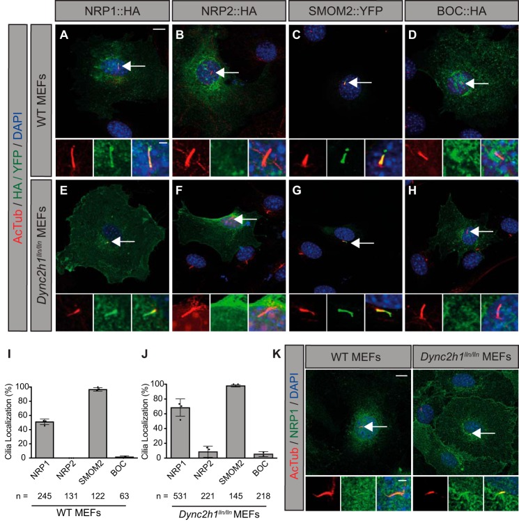
Figure 6.
NRP1, but not NRP2, localizes to primary cilia in HH-responsive fibroblasts. A–H, antibody detection of HA/YFP (green) and primary cilia (red, AcTub) in WT (top) and Dynein-mutant (Dync2h1lln/lln) MEFs (bottom). Dync2h1lln/lln MEFs exhibit impaired retrograde transport out of primary cilia. Arrows indicate the location of primary cilia. Insets show higher-magnification views of primary cilia in individual (left and center) and merged (right) channels. DAPI indicates nuclei (blue). I and J, quantitation of data represented in images from WT (left) and Dync2h1lln/lln (right) MEFs, reported as mean ± S.D., with scatterplot values indicating averages of three independent experiments and the total number of cells analyzed listed below each bar. Data are reported from at least three independent experiments. K, antibody detection of endogenous NRP1 in WT (left panel) and Dync2h1lln/lln (right panel) MEFs. Scale bar = 10 μm, inset scale bar = 1 μm.

