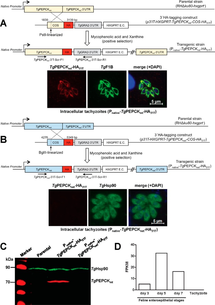
Figure 2.
TgPEPCKmt (TgPEPCK1) localizes in the mitochondrion, whereas TgPEPCKnet (TgPEPCK2) is not expressed in the tachyzoite stage of T. gondii. A, 3′-insertional tagging of the TgPEPCKmt gene with a HA tag in tachyzoites and detection of TgPEPCKmt-HA3′IT protein by immunofluorescence. The PstI-digested plasmid construct with a COS targeting the 3′-end of the TgPEPCKmt gene was transfected into the RHΔku80-hxgprt− strain. Tachyzoites encoding TgPEPCKmt-HA3′IT under the control of its native promoter and TgGRA2 3′-UTR were selected with the indicated drugs, screened by genomic PCR using TgPEPCKmt-3′IT-Scr-F1/R1 primers, and verified by sequencing. Intracellular parasites (24-h infection) were immunostained with α-HA and α-TgF1B antibodies to determine the subcellular location of TgPEPCKmt-HA3′IT and treated with DAPI to visualize the cell nuclei. B, genomic tagging of the TgPEPCKnet gene and fluorescent detection of TgPEPCKnet-HA3′IT in tachyzoites. Stable transgenic parasites were generated and immunostained with α-HA and α-TgHsp90 antibodies, as stated in A. C, immunoblotting showing the natural expression of TgPEPCKmt-HA3′IT, TgPEPCKnet-HA3′IT, and TgHsp90 in transgenic tachyzoites from A and B. Extracellular parasites (107) of the indicated strains were subjected to protein isolation followed by SDS-PAGE and immunostaining using α-HA and α-TgHsp90 (loading control) antibodies. Parental strain served as a negative control for α-HA staining. D, comparative levels of TgPEPCKnet RNA in the tachyzoite and merozoite stages of T. gondii. The graph is reproduced from the parasite database (www.ToxoDB.org)5 based on previous work of A. B. Hehl et al. (20). Tachyzoites were grown in vitro; merozoites were isolated from enterocytes of the cyst-infected cats on specified days. The y axis denotes transcript levels of fragments per kilobase of exon model per million mapped reads (FPKM), as measured by standard paired-end Illumina sequencing of the parasite mRNA.

