Figure 3.
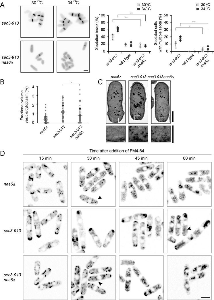
Deletion of nas6 restores Sec3-913 function in endocytosis and exocytosis. A, calcofluor staining of sec3-913 and sec3-913nas6Δ cells at 30 °C and 34 °C (left panel). The percentages of septated cells (septation index) and of septated cells with multiple septa were determined. The error bars indicate the standard deviation (n = 4). *, p < 0.05; **, p < 0.01; ***, p < 0.001. Scale bar = 5 μm. B, by electron microscopy, the fractional volume of vesicles in the cytoplasm of the indicated cells grown at 34 °C was determined. Whiskers mark the standard deviation (n > 202). The results are representative of three independent experiments. *, p < 0.05. C, transmission electron microscopy images of nas6Δ, sec3-913, and sec3-913nas6Δ cells at 34 °C. Higher magnifications of the boxed areas show fewer accumulated vesicles in sec3-913nas6Δ than in sec3-913. Scale bars = 2 μm for whole cells and 1 μm for the enlargement. D, the indicated strains were grown at 36 °C with FM4-64 dye and analyzed at regular intervals up to 1 h following addition of the dye. The accumulation of FM4-64 at the tips of sec3-913 cells and its absence from endosomes are indicative of a defect in endocytosis. Note how this phenotype is reversed in the sec3-913nas6Δ double mutant (arrowheads). Scale bar = 5 μm.

