Figure 2.
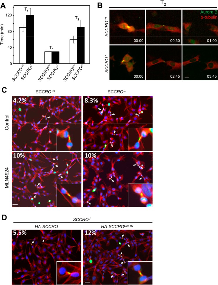
Depletion of SCCRO in MEFs delays abscission. A, comparison of duration of metaphase and cytokinesis between SCCRO+/+ (white columns) and SCCRO−/− (black columns) MEFs. Note that, although T1 is essentially the same for both cells, SCCRO−/− MEFs have a T2 ∼50% longer than that in SCCRO+/+ MEFs. B, long-term imaging of cell division of SCCRO+/+ and SCCRO−/− MEFs stably expressing Aurora B-EGFP and mCherry–α-tubulin. Compared with SCCRO+/+ MEFs (top row), SCCRO−/− MEFs show delayed abscission (bottom row). Scale bar = 5 μm. C, immunofluorescence using anti-Aurora B (green), anti-α-tubulin (red), and DAPI (blue), showing an increased percentage of midbody cells in SCCRO−/− MEFs compared with SCCRO+/+ MEFs (top row). Treatment with MLN4924 (1 μm for 2 h) increased midbody cells in SCCRO+/+ MEFs to a level similar to that in SCCRO−/− MEFs (bottom row). Insets show a close-up view of midbody cells in each case. The numbers are the percentages of midbody cells. Scale bar = 20 μm. D, the increased percentage of midbody cells seen in SCCRO−/− MEFs was rescued by retroviral introduction of HA-SCCRO but not HA-SCCROD241N.

