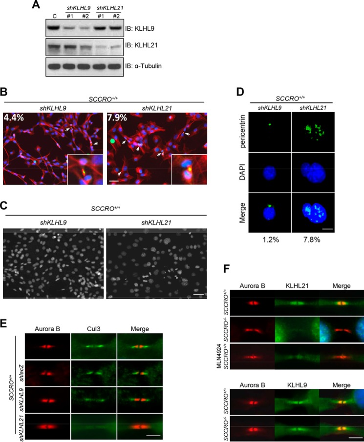
Figure 4.
SCCRO regulates abscission through the Cul3KLHL21 complex. A, Western blot analysis of lysates from SCCRO+/+ MEFs treated with shRNA against KLHL9 or KLHL21 as indicated and probed with KLHL9, KLHL21, and α-tubulin antibodies. C, lacZ shRNA knockdown control; IB, immunoblot. B, KLHL9 (left panel) and KLHL21 (right panel) shRNA-treated SCCRO+/+ MEFs stained for Aurora B (green), α-tubulin (red), and nuclei (blue). KLHL21 shRNA-treated cells show increased levels of midbody cells. Insets, close-up view of midbody cells. The percentages of midbody cells are included inside the images. Scale bar = 20 μm. C, DAPI staining, showing an increased percentage of polyploidy cells in SCCRO+/+ MEFs with KLHL21 knockdown (right panel) compared with those with KLHL9 knockdown (left panel). Scale bar = 20 μm. D, immunofluorescence analysis using anti-pericentrin, showing supernumerary centrosomes in SCCRO+/+ MEFs with KLHL21 knockdown but not in those with KLHL9 knockdown. Scale bar = 5 μm. E, immunostaining for Cul3 and Aurora B, showing an absence of Cul3 from the midbody in SCCRO+/+ MEFs with KLHL21 knockdown (shKLHL21) but not in SCCRO+/+ MEFs with KLHL9 knockdown (shKLHL9) or lacZ knockdown controls (shlacZ). Scale bar = 5 μm. F, immunostaining for KLHL21 and Aurora B, showing an absence of KLHL21 in the midbody of SCCRO−/− MEFs and MLN4924-treated SCCRO+/+ MEFs compared with SCCRO+/+ MEFs (top panel). Note that there is no difference in KLHL9 localization between SCCRO+/+ and SCCRO−/− MEFs (bottom panel).

