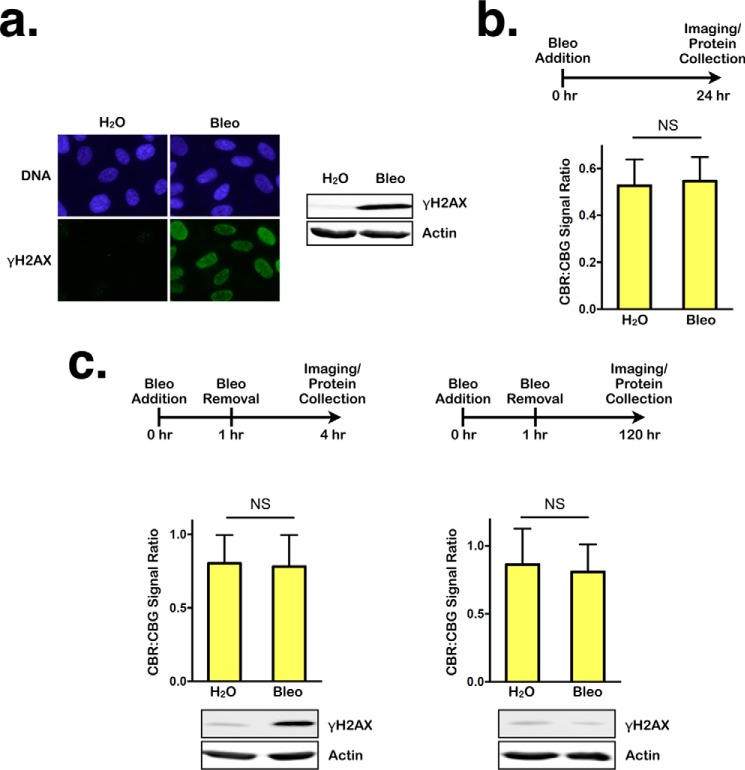
Figure 2.
Transient DNA damage does not affect NMD. a, left panel, representative images of immunofluorescence staining for DNA or γH2AX in RPE1 cells after 24-h treatment with either H2O or bleomycin (Bleo). Right panel, Western blot showing γH2AX levels in lysates collected after 24-h treatment with either H2O or bleomycin. b, ratios of CBR:CBG bioluminescence signals in RPE1 reporter cells after 24-h treatment with either H2O or bleomycin. Data represent the mean ± S.D. of three independent experiments. NS, p > 0.05 (paired t test). c, left panel, ratios of CBR:CBG bioluminescence signals (top panel) and γH2AX levels (bottom panel) in RPE1 reporter cells after 1-h treatment with either H2O or bleomycin, followed by 3-h recovery. Data represent the mean ± S.D. of three independent experiments. NS, p > 0.05 (paired t test). Right panel, ratios of CBR:CBG bioluminescence (top panel) and γH2AX levels (bottom panel) signals in RPE1 reporter cells after 1-h treatment with either H2O or bleomycin followed by 120-h recovery. Data represent the mean ± S.D. of three independent experiments. NS, p > 0.05 (paired t test).

