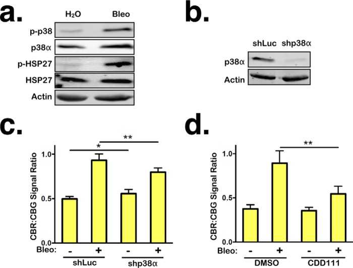
Figure 4.
NMD inhibition by persistent DNA damage is mediated in part by p38α. a, levels of total p38α and phosphorylated p38 and HSP27 in RPE1 cells treated with either H2O or bleomycin (Bleo) for 24 h, followed by 96-h recovery. b, shRNA-mediated knockdown of p38α in RPE1 cells. c, ratios of CBR:CBG bioluminescence signals in control and p38α knockdown RPE1 reporter cells after 24-h treatment with either H2O or bleomycin, followed by 96-h recovery. Data represent the mean ± S.D. of four independent experiments. **, p ≤ 0.01 (paired t test). d, ratios of CBR:CBG bioluminescence signals in DMSO- or CDD111-treated RPE1 reporter cells after 24-h treatment with either H2O or bleomycin, followed by 96-h recovery. Data represent the mean ± S.D. of three independent experiments. *, p < 0.5; **, p ≤ 0.01 (paired t test).

